
Understanding the Types of Medical Imaging Technology
 If you’re injured in an accident or experience unknown medical problems, it’s fairly common for your primary care doctor to order some type of medical imaging in an effort to accurately diagnose your condition. Medical imaging centers offer a wide range of diagnostic tools that allow medical professionals to look inside your body without an invasive surgical procedure. A variety of different imaging tests may be scheduled for you:
- X-ray—Invented over 125 years ago, X-ray technology revolutionized modern medicine. X-rays use a type of ionizing radiation, invisible to the naked eye, that passes through your body. The denser parts of your body show up as white areas on the image produced. X-rays are most effective at producing images of bones and teeth.
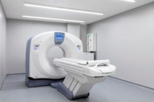
- CT—Computed tomography (known as CT or CAT) scans, like X-rays, also use ionizing radiation but can produce images with a significantly higher level of detail, including 360-degree views of body structures, including organs, bones, and blood vessels. CT scans can be extremely useful in the diagnosis and treatment of bone or organ injury, stroke, cancer, and vascular injury or illness.
- MRI—Magnetic resonance imaging (MRI) uses radio waves, created by a magnet, that interact with protons in your body to create detailed images. An MRI can show images of soft tissue, blood vessels, and nerves, as well as bones and teeth. Magnetic resonance cholangiopancreatography (MRCP) is a type of MRI that employs radio waves produced by a magnetic field to create computerized images of the pancreas, pancreatic duct, gallbladder, bile ducts, and liver.
- Fluoroscopy—Think of fluoroscopy as an X-ray movie. It uses a continuous X-ray beam to produce a screen image. It can be used to diagnose urinary problems, joint issues, blood vessel and organ illnesses, and challenges with the gastrointestinal tract.
- Arthrogram—Arthrography involves injecting a contrast dye into a joint. X-rays are then taken to create pictures of the joint. It’s commonly used to treat potential arthritic pain in knees, shoulders, or hips. An arthrogram may also use CT or MRI technology.
- Myelogram—This procedure uses contrast dye and either X-rays or a CT scan to diagnose medical problems in your spinal canal, including issues with nerve roots and tissue.
- Ultrasound—An ultrasound employs high frequency sound waves to create images of internal organs and body structures. The process takes up to an hour and is frequently used to monitor pregnancy or diagnose blood flow issues, gallbladder problems, lumps, or tumors. The picture generated by an ultrasound is called a sonogram.
- PET scan—Positron emission tomography (PET) uses radioactive drugs, known as tracers, and scanning technology to look at how your organs and tissues function. It’s typically a much longer process than X-rays, CT scans, and MRIs. PET scans are common in the treatment of cancer, heart disease, epilepsy, and Parkinson’s.
Contact InjuredCare to Connect with an Experienced Medical Imaging Professional
To get connected with the right professional to conduct a medical imaging test, contact us online or call our offices today at 866-952-7045.
